
HIGHLIGHTS
- Cyclic, high-throughput FISH-based imaging
- From lab setup to user-friendly, walk-away instrument
- Multi-disciplinary team for integrated design
- Delivery of alpha and beta prototypes within two years
- Timely delivery of system despite global travel restrictions
unlocking the potential of spatial omics.
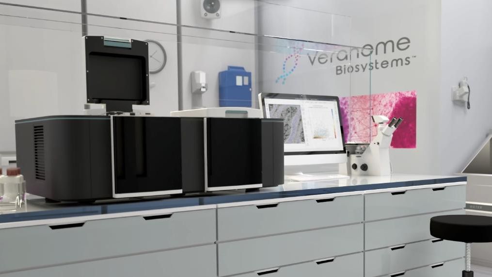
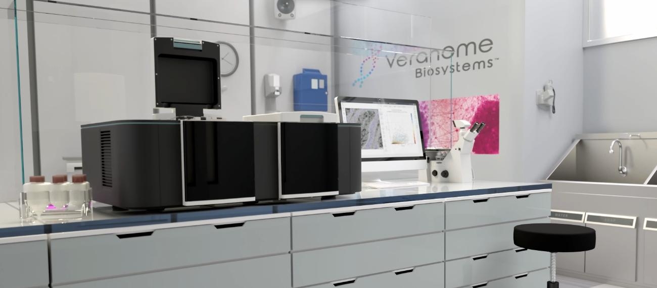
Spatial genomics is a rapidly evolving field that combines traditional genomics techniques with spatial information. This helps advance scientific understanding of the organization and function of cells and tissues within their native context, enabling a wide range of applications across various biological disciplines. To visualize the highly multiplexed and spatial detection of RNA, a specialized system is needed. This system must be capable of fluorescently labeling and imaging samples in a cyclic manner and with high throughput. The customer, Veranome Biosystems, an Applied Materials company, requested to turn their initial lab setup based on standard equipment into an easy to use ‘walk away’ automated product, including multiple optimizations.
next generation spatial omics
engineering challenges for high-precision RNA imaging.
The technical challenges posed were:
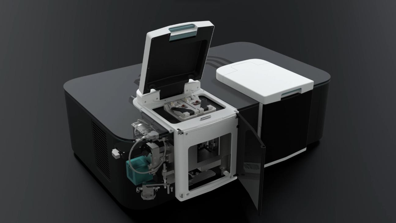
- Custom development of an integrated laser-based oil-immersion microscope in a vibration damped configuration.
- The safe autofocus design in order to enable very small working distances while preventing crashing the objective into the sample.
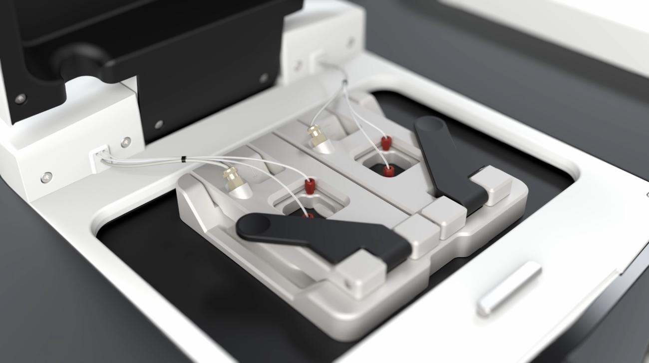
- The design of a user-friendly leakage-free flow cell. The flow cell module positioned on top of the optical module, had to be stable and robust for internal and external disturbances enabling ultra-high-resolution images needed for the visualization of mRNA.
Distance does not have to be a challenge
accelerating time to market.
The development had to be done in a short timeline with just two years for the de-risking steps as well as the alpha and beta design iterations in order to start series production. Meanwhile, COVID travel restrictions proved another challenge, with the customer seated in both Asia and the USA requiring all customer communication to be remote.

integrated system for transcriptomics and gene expression mapping.
The resulting design is a tabletop system including easy to use flow cells for sample loading, a vibration damped oil immersion microscope and a fluidic system. The optical module consists of an imaging & illumination path and autofocus to produce high resolution, highly homogeneous stitched images. A fluidics module selects and controls 34 reagents flowing through the flow cell containing the tissue sample fixed on a microscope slice. A flow cell module can move with high accuracy and precision to scan different areas of the sample.
"Accelerate spatial genomics research by developing compact, commercial-grade and user-friendly tabletop systems for spatial transcriptomics with us."